

Sleep apnea: Retraction of the mandible compresses the airway

The operating room at Feng Hua Joint Clinic is equipped with surgical lights and anesthesia machines.
Comparison of facial profile and airway before and after 3D customized orthognathic surgery for patients with receding jaw
Preoperative symptomssurgical plan
Retruded jaw, no chin affecting airway space, severe sleep apnea3D customized orthognathic surgery

postoperative changes

Dr. Xie Mingji | Sleep Apnea | Orthognathic Surgery

Diagnosis and treatment plan and postoperative changes

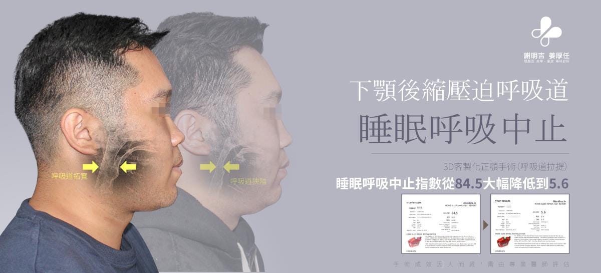
The patient was diagnosed with severe sleep apnea (AHI index as high as 84.5, and by definition an AHI index of ≥30 is considered severe sleep apnea). From his computed tomography before surgery, it was shown that he belonged to type 2 bones. (That is, a receding jaw and no chin) This is a typical face shape for people with sleep apnea.

A retracted chin compresses the airway space, causing the airway to become narrow. After airway lift (orthognathic surgery), his AHI index was significantly reduced from 84.5 to 5.6, and the minimum blood oxygen concentration was significantly increased from 50% to 90%.

In the current literature, successful treatment is defined as a reduction in the AHI index below 10 or a 50% reduction in the AHI index before and after surgery.

Sleep monitoring reports comparing pre- and post-operative results showed that the AHI decreased from 84.5 and the lowest blood oxygen saturation increased from 501 TP3T to 901 TP3T.

As we can see from the photos, the orthognathic surgery significantly widened his airway and also improved his appearance, making his jaw more proportional.

We are so happy that he can now breathe easier, sleep better, and face a better life.

Comparison of the facial airway before and after orthognathic surgery; postoperative airway widening.

Appearance changes

Frontal appearance comparison before and after orthognathic surgery for patients with mandibular retrusion
Comparison of 45-degree bevel appearance before and after orthognathic surgery
Comparison of chin and jaw contours before and after orthognathic surgery
Comparison of side-view photos before and after surgery

Comparison of tomography before and after surgery

3D computed tomography before and after orthognathic surgery showed a significant increase in airway space postoperatively.

Recovery progress

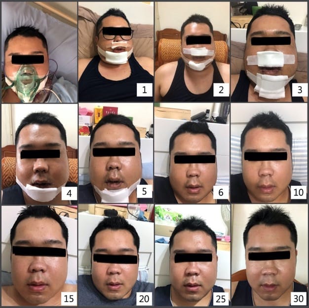
A record of facial recovery from day 1 to day 30 after orthognathic surgery.

Warm reminder

  1. Please follow the instructions of the doctor and nursing staff, take medicine regularly, pay attention to dietary taboos and keep the wound clean.
  2. There may be complications such as bleeding, infection, inflammation, etc. after surgery. The degree will vary according to individual physical differences, so careful care is required. If the above situations occur, please contact us as soon as possible.
  3. It is normal for 3 to 6 months after surgery to experience temporary slow sensation and unnatural expressions at the surgical site. Through independent massage and facial expression exercises, most patients will gradually improve within 6 to 12 months after surgery.
  4. Anesthesia has certain risks, which are related to the patient's own physical condition. Patients must honestly and clearly report their health status and treatment history to the treating physician and anesthesiologist, and do not hide anything.
  5. It is recommended to communicate with the doctor before surgery and clearly express treatment needs. However, the aesthetic appearance after surgery is subjective and should not be used as a basis for judgment of surgical results.
  6. The results of surgery vary from person to person and need to be evaluated by a professional physician. Case pictures are publicly used with the authorization of the parties involved as auxiliary explanations of diagnosis and treatment, health education and medical knowledge sharing, and are for reference only.