



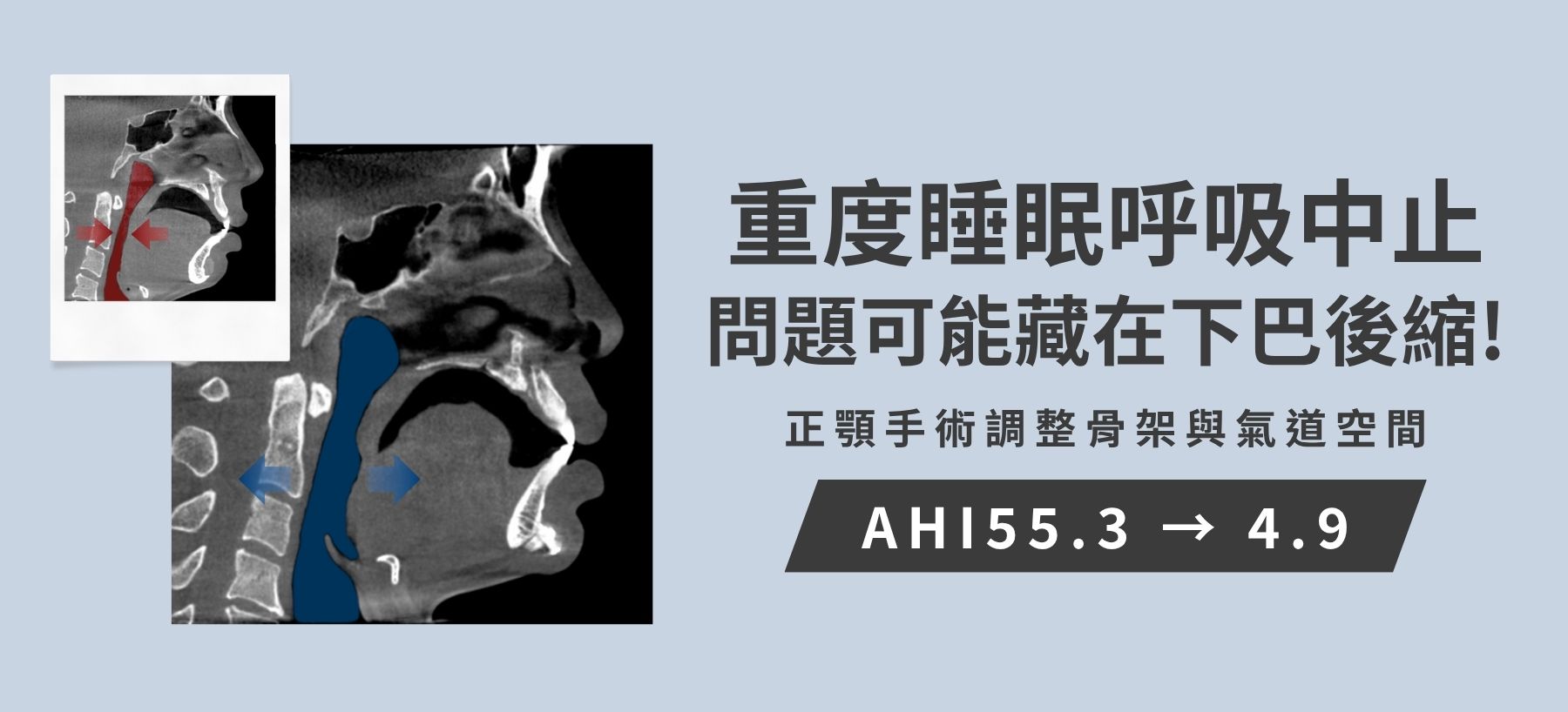
| Preoperative symptoms | surgical plan |
| Retrusion of the upper and lower jawbones Severe obstructive sleep apnea The hyoid bone is abnormally low in position. | 3D Customized Orthognathic Surgery (Bimaxillary Rotation and Advancement Surgery) Elevation and anterior displacement of the low hyoid bone Contour Reconstruction |
In the clinical management of patients with moderate to severe obstructive sleep apnea (OSA), if the cause is mainly due to craniofacial skeletal deficiency, and in cases where compliance with traditional soft tissue surgery or positive airway pressure (CPAP) is poor, maxillomandibular advancement (MMA) often plays a key role in skeletal remodeling.
This article shares a case of a 37-year-old male with severe OSA and significant daytime sleepiness (EDS) who underwent interdisciplinary referral:
I. Preoperative assessment and analysis of anatomical abnormalities
- Sleep physiology findings: Preoperative home sleep testing (HST) showed severe OSA, with an AHI as high as 55.3 beats/hour (62.2 beats/hour in supine position), a 3% ODI of 55.4 beats/hour, a minimum oxygen saturation (Min SpO2) dropping to 77.0%, and a cumulative hypoxic time of 46.0 minutes (accounting for 11.1% of total sleep time) with oxygen saturation below 88%.
- Craniofacial and respiratory tract computed tomography (CT) assessment:
- The maxillary and mandible complexes show significant retrusion, resulting in severe narrowing of the anteroposterior diameter of the oropharynx and hypopharynx.
- The hyoid bone is abnormally low in position, and the muscles at the base of the tongue lack sufficient bony attachment to pull forward, causing the base of the tongue to collapse and obstruct the posterior airway when lying flat.
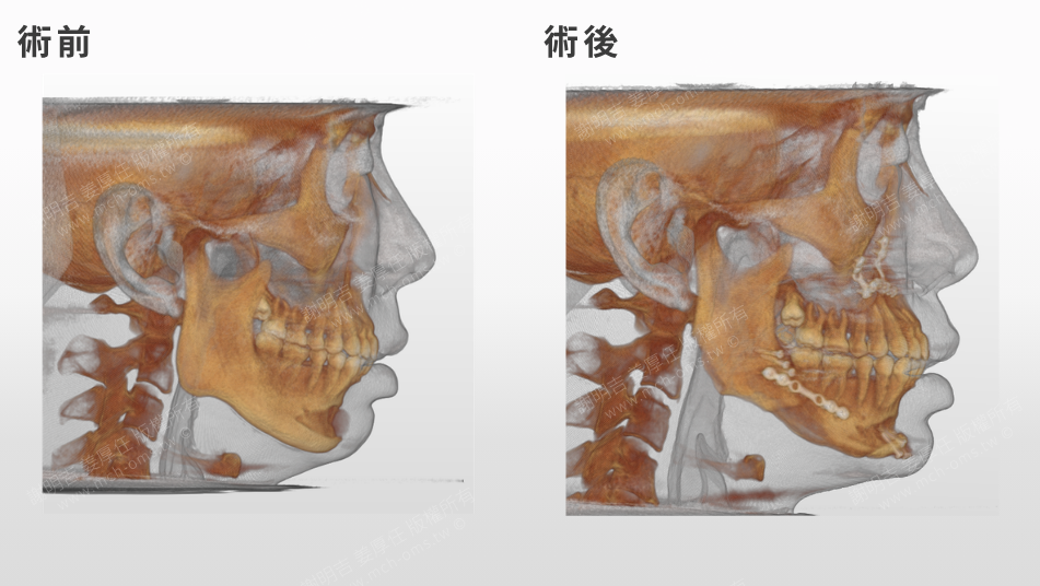
- The patient underwent a large silicone chin implant elsewhere several years ago. CT scans showed that the implant caused local cortical bone compression and erosion, but did not resolve the physiological structural limitations of the retraction of the genioglossus muscle and suprahyoid muscles.
II. Surgical Planning and Execution Strategies
- Counter-Clockwise MMA: Through Le Fort I osteotomy and bilateral mandibular branch sagittal splitting (BSSO), the occlusal plane is rotated in a CCW manner, achieving full forward displacement of the mandibular body and lingual base while controlling the vertical height of the mid-lower face and the projection angle of the anterior teeth.
- Silicone prosthesis removal combined with autologous bone advancement genioplasty: The silicone prosthesis is completely removed during the operation and replaced with anterior osteotomy and fixation; this can pull the submental muscles and genioglossus muscles forward, causing the tongue base to move forward away from the posterior pharyngeal wall.
III. Postoperative CT Imaging and Structural Interpretation
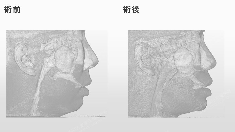
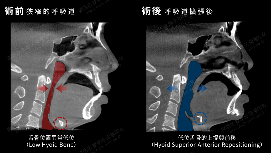
- Posterior airway volume expansion (PAS Expansion): Postoperative sagittal section and 3D airway reconstruction show that the minimum anteroposterior diameter of the oropharynx and hypopharynx is expanded by multiples, and the originally collapsed posterior pharyngeal wall space, which was in the form of a narrow slit, is restored to a spacious cylindrical tube.
- Hyoid Superior-Anterior Repositioning: As the suprahyoid muscles and mandibular body move forward, the hyoid bone is moved to an anterior and superior anatomical position, significantly increasing the supporting tension of the pharyngeal lateral wall and the base of the tongue.
- Contouring and proportion reconstruction: Improves the skeletal contours of a receding chin and blunted mandibular angle, reduces the anatomical abruptness caused by implants, and balances function and facial aesthetics.
IV. Comparative Analysis of Postoperative Sleep Monitoring (HST) Data
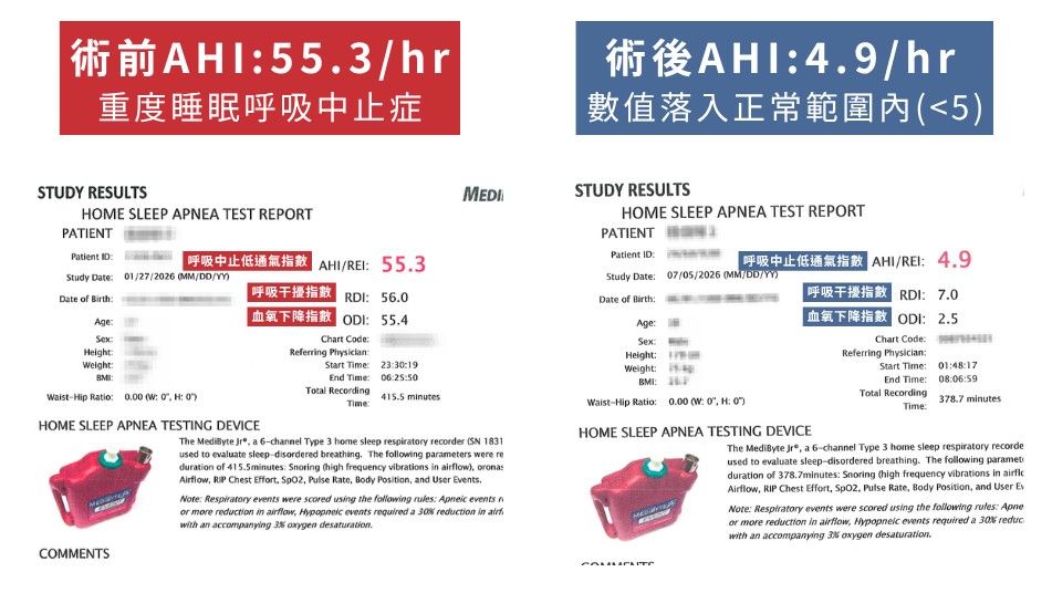
Postoperative sleep tracking reports showed improvements in physiological indicators:
- AHI/REI (Apnea-Hypopnea Index): decreased from 55.3 breaths/hour to 4.9 breaths/hour (6.6 breaths/hour in supine position), falling into the normal range (<5).
- 3%ODI (number of times blood oxygen drops by ≥ 3% per hour): decreased from 55.4 times/hour to 2.5 times/hour.
- RDI (Relative Oxygen Degradation Index): decreased from 56.0 to 7.0 beats/hour (reaching the mild borderline).
- Blood oxygen performance: The lowest blood oxygen level increased from 77.01 TP3T to 91.01 TP3T; more significantly, the time when blood oxygen was below 881 TP3T decreased from 46.0 minutes to 0 minutes (0.01 TP3T), and the blood oxygen concentration was maintained above 901 TP3T throughout the night during sleep with a total blood oxygen level of 1001 TP3T.
- Clinical symptoms: The patient reported relief from daytime sleepiness, and the headache and shortness of breath upon waking no longer occurred. Daytime mental alertness and work concentration improved.
[Clinical Summary]
For severe OSA with skeletal retraction, MMA (meditative retraction) not only expands the space behind the soft palate, but also pulls the entire hyoid apparatus by moving the skeleton forward. We welcome exchanges and referrals among specialists to evaluate and collaborate on providing treatment options that combine structural reconstruction for patients with refractory sleep apnea.