

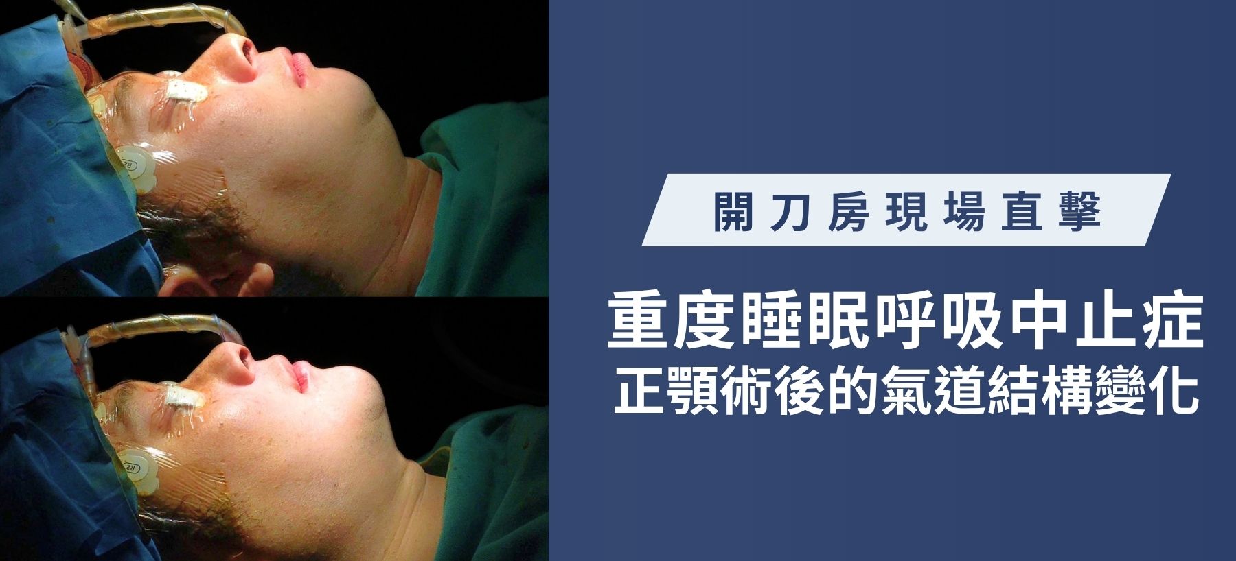
Live from the operating room: Mandibular retrusion combined with severe sleep apnea, airway structural changes during MMA orthognathic surgery!

The operating room at Feng Hua Joint Clinic is equipped with surgical lights and anesthesia machines.
Changes in airway structure after orthognathic surgery for severe sleep apnea

Many patients with severe sleep apnea (OSA) often ask me, "Dr. Hsieh, can orthognathic surgery really improve breathing?"

Today's photos are the most authentic answer from inside the operating room.

This patient had long suffered from "receding mandible." Structurally, when the mandible recedes excessively, it directly compresses the airway space behind, causing the tongue to fall back and the airway to narrow severely when lying down to sleep, eventually developing into severe sleep apnea. This not only affects sleep quality but is also a hidden killer of cardiovascular health.

To address this fundamental structural problem, we employed MMA (Maxillomandibular Advancement, orthognathic surgery).
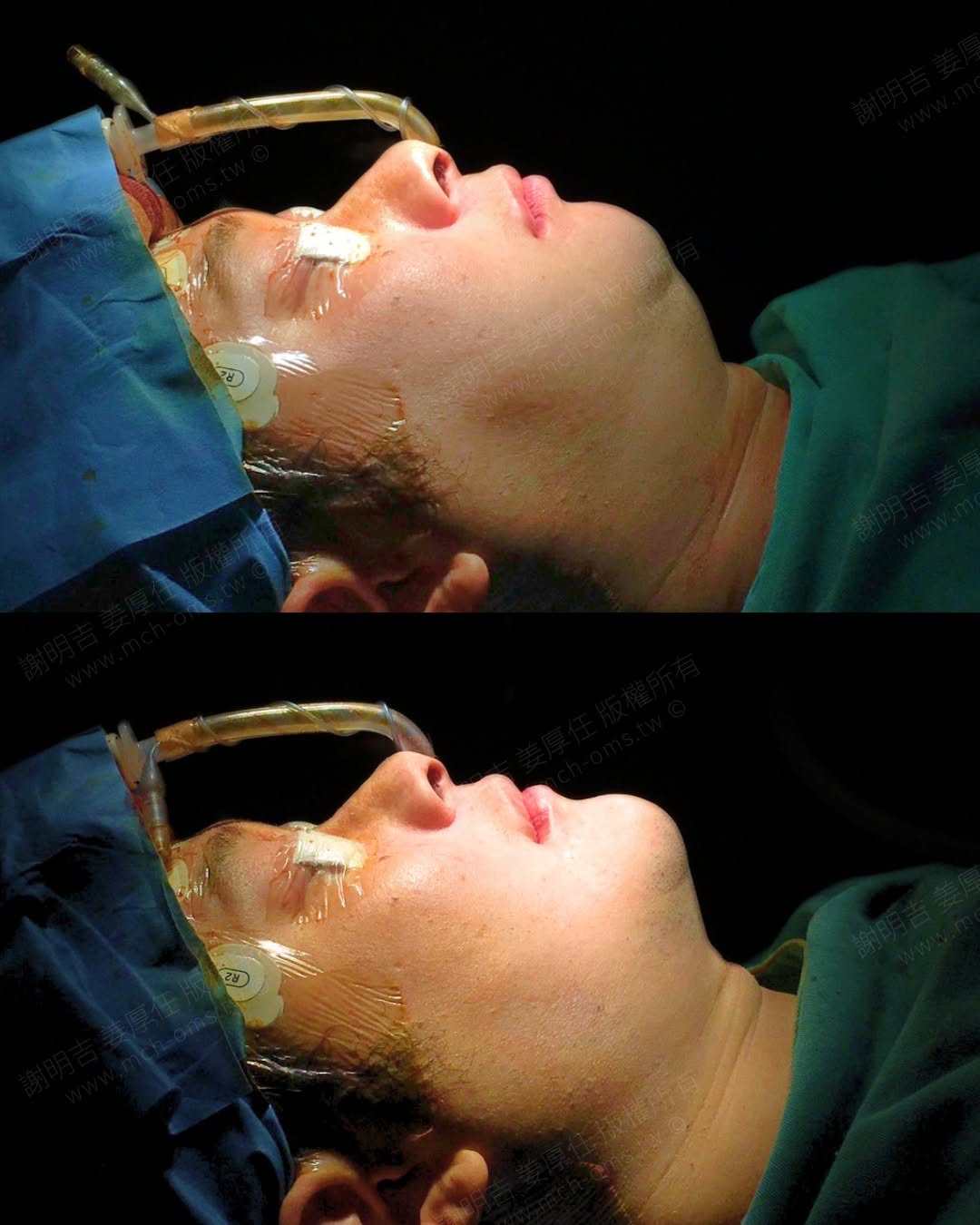
Side profile comparison of a patient with mandibular retrusion and severe sleep apnea (OSA) before and after MMA orthognathic surgery.

By comparing the before-and-after images of a patient in the operating room with a breathing tube still in place, the structural changes can be clearly seen:

  • 1. The chin moves forward immediately: The previously retracted jawbone is precisely brought to the ideal skeletal position.
  • 2. Changes in neck lines: As the bones move forward, the previously loose or compressed soft tissues of the neck change with the movement of the bones, making the profile lines tighter and smoother.

The change in appearance is just an "added value" of orthognathic surgery; our core purpose is to "improve airway space".

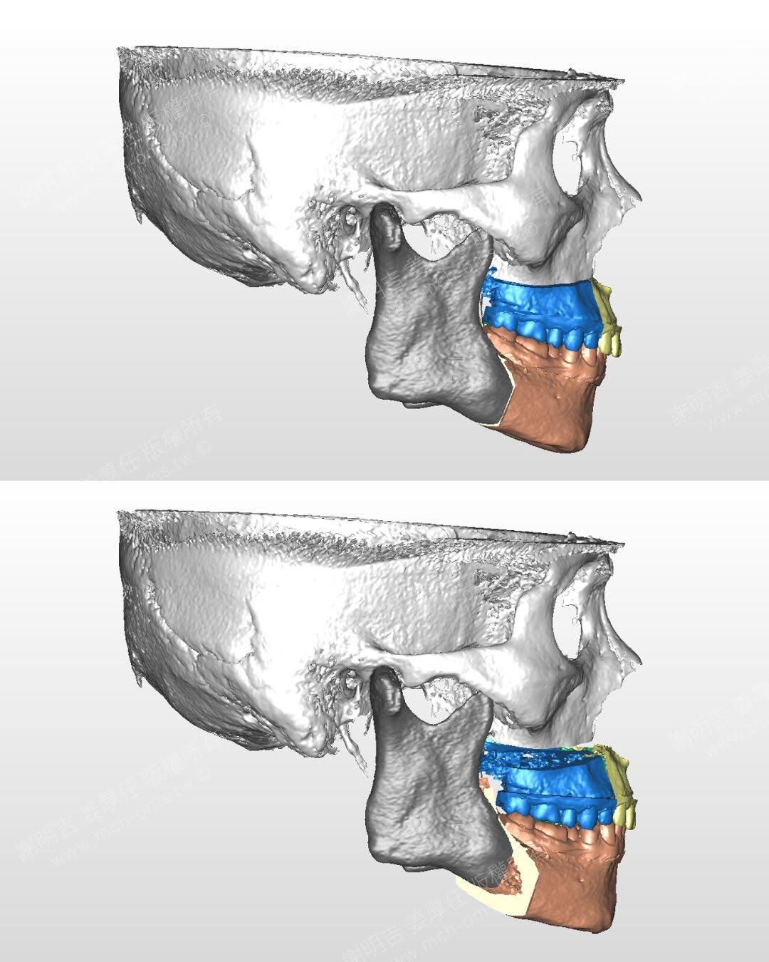
Along with the "3D digital surgical plan" in the post, we precisely simulated the angle at which the upper and lower jaws needed to be moved forward and the position of the bone screws before the operation. And for the most crucial evidence, please see the final comparison of the "postoperative CT computed tomography scan":

  • Preoperative (red area): The airway is severely "narrowed" and airflow is obstructed.
  • Postoperative (blue area): The entire upper airway space "increased" compared to before the operation.
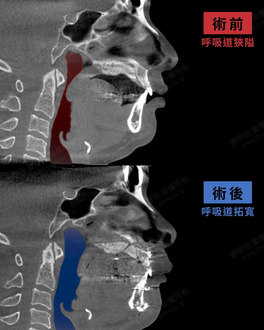
Computed tomography airway space comparison before and after MMA orthognathic surgery in patients with mandibular retrusion and severe sleep apnea (OSA).

3D jawbone position comparison before and after MMA orthognathic surgery in patients with mandibular retrusion and severe sleep apnea (OSA).

With the change in structure and the creation of space, patients can immediately feel the ease of "breathing deeply" the moment they wake up from anesthesia and are extubated.

This is the most fascinating and medically valuable core of craniofacial orthognathic surgery—improving airway space through adjustments to the position of the jawbone.

Warm reminder

  1. Please follow the instructions of the doctor and nursing staff, take medicine regularly, pay attention to dietary taboos and keep the wound clean.
  2. There may be complications such as bleeding, infection, inflammation, etc. after surgery. The degree will vary according to individual physical differences, so careful care is required. If the above situations occur, please contact us as soon as possible.
  3. It is normal for 3 to 6 months after surgery to experience temporary slow sensation and unnatural expressions at the surgical site. Through independent massage and facial expression exercises, most patients will gradually improve within 6 to 12 months after surgery.
  4. Anesthesia has certain risks, which are related to the patient's own physical condition. Patients must honestly and clearly report their health status and treatment history to the treating physician and anesthesiologist, and do not hide anything.
  5. It is recommended to communicate with the doctor before surgery and clearly express treatment needs. However, the aesthetic appearance after surgery is subjective and should not be used as a basis for judgment of surgical results.
  6. The results of surgery vary from person to person and need to be evaluated by a professional physician. Case pictures are publicly used with the authorization of the parties involved as auxiliary explanations of diagnosis and treatment, health education and medical knowledge sharing, and are for reference only.