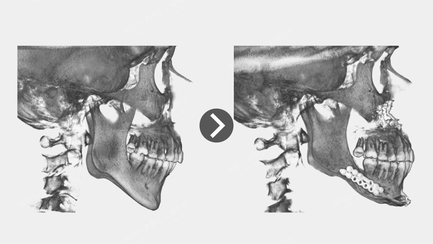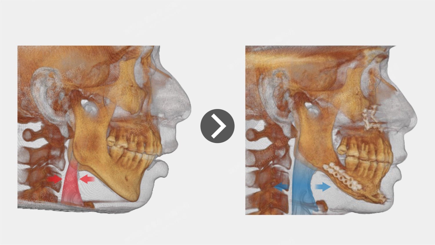




| Preoperative symptoms | surgical plan |
| Severe nocturnal snoring and daytime sleepiness, moderate sleep apnea (AHI 17.3) | Performing a counterclockwise rotational double jaw advancement surgery to move the upper and lower jaws and hyoid bones forward to widen the airway |
Diagnosis and treatment plan and postoperative changes
The patient complained of severe nocturnal snoring and daytime sleepiness, which impacted his quality of life. Multiple sleep studies revealed moderate obstructive sleep apnea (AHI = 17.3), with the lowest blood oxygen concentration during sleep dropping to 831 TP3T, indicating significant ventilation impairment.
This patient presented with complaints of severe snoring during sleep and excessive daytime sleepiness, significantly affecting quality of life. Polysomnography revealed moderate obstructive sleep apnea (OSA) with an apnea–hypopnea index (AHI) of 17.3, and a minimum oxygen saturation of 83% during sleep, indicating substantial ventilatory compromise.
CT scans revealed a significant narrowing of the patient's posterior airway, primarily due to underdeveloped maxillary and mandibular bones, which failed to grow forward properly. Maxillary retraction and airway collapse caused the loose septum to vibrate in the confined space, resulting in noticeable snoring. Mandibular retraction, in turn, led to a posterior tongue tilt, insufficient tongue muscle tone, and a low hyoid bone position, becoming the primary structural factor in obstructive sleep apnea.
Computed tomography (CT) imaging demonstrated a markedly narrowed posterior airway space, primarily due to underdeveloped maxillary and mandibular growth. The retruded maxilla, combined with airway collapse, allowed the relaxed uvula to vibrate within the constricted space, producing loud snoring. The retrognathic mandible further contributed to posterior tongue displacement, reduced tongue muscle tone, and a low-positioned hyoid bone—key anatomical factors in the pathogenesis of OSA.
In view of the above anatomical and functional issues, we planCounter-Clockwise Rotation Maxillomandibular Advancement (CCWR-MMA).
There are three central ideas in the entire surgical design:
1. The forward movement of the maxillary bone effectively tightens the soft palate muscles, pulling the suspensory part forward and away from the posterior pharyngeal wall, thus reducing snoring.
2. The forward movement of the mandible pulls the tongue root, causing the tongue to move forward and reduce the backward movement of the tongue root.
3. At the same time, the hyoid bone is also pulled to the front and upper position, away from the posterior wall of the airway, significantly expanding the pharyngeal cavity space and improving ventilation efficiency.
To address these structural and functional issues, we planned a counter-clockwise rotation maxillomandibular advancement (CCWR-MMA) procedure. This surgical approach included:
- Maxillary advancement, which tightened the soft palate musculature and repositioned the uvula anteriorly, away from the posterior pharyngeal wall, thereby reducing snoring;
- Mandibular advancement, which, through its anatomical connection to the tongue base, repositioned the tongue anteriorly, minimizing posterior collapse;
- Additionally, the hyoid bone was drawn forward and upward, distancing it from the posterior airway wall and significantly expanding the pharyngeal space to improve airflow.
Preoperative and postoperative CT images clearly showed that after the bone position was adjusted, the soft tissue tension was increased, the airway space was widened, and the structural improvement was clear.
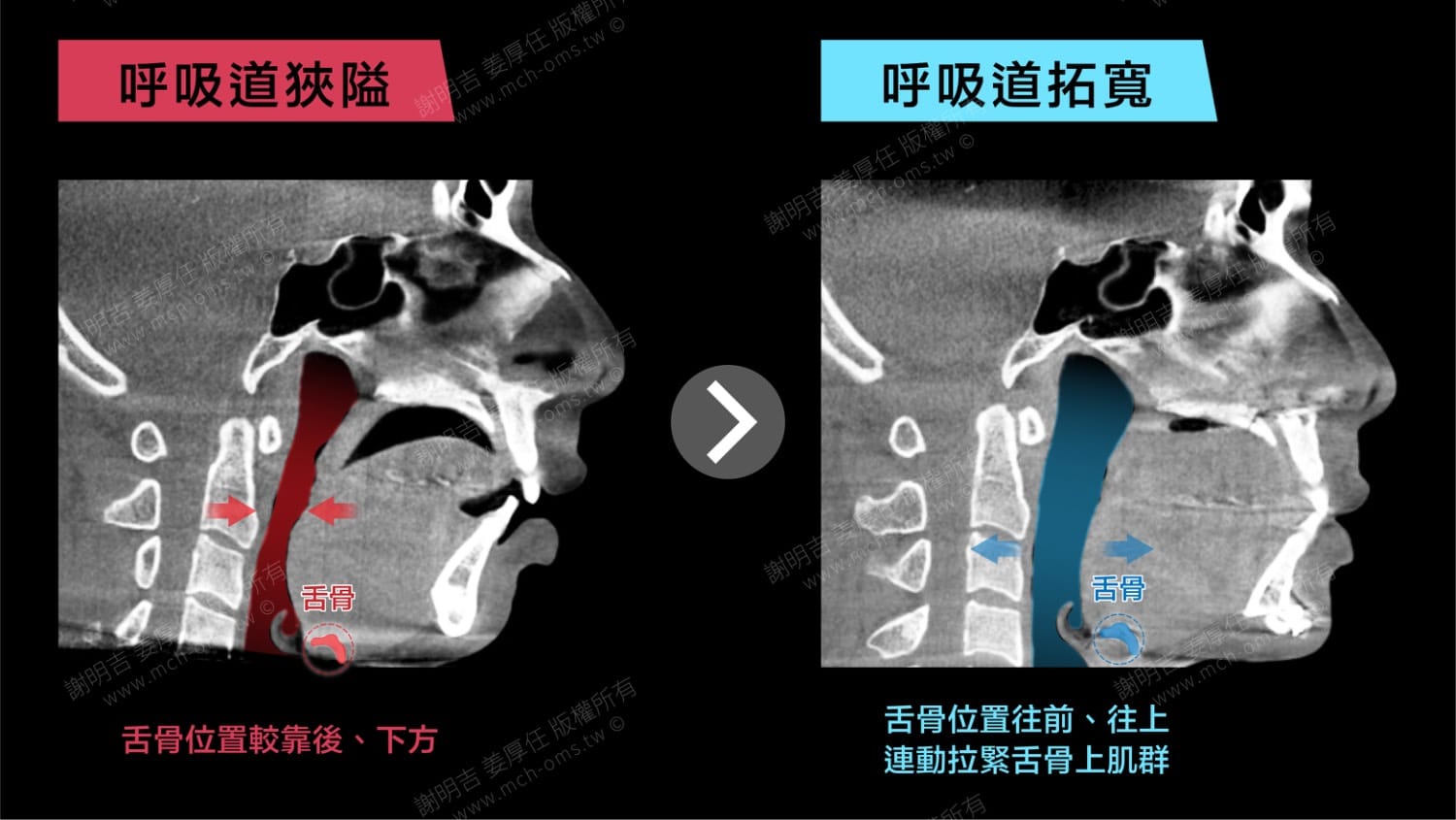
Postoperative sleep reports showed an AHI of 6.3, with the lowest blood oxygen concentration increasing to 891 TP3T. The patient reported significant improvements in sleep quality, brighter daytime mental state, and improved overall quality of life.
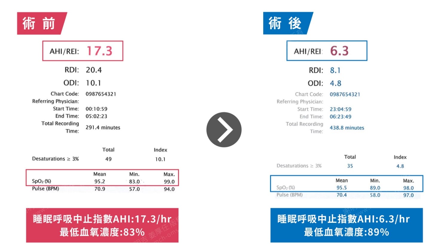
Pre- and postoperative CT scans clearly demonstrated how skeletal repositioning led to soft tissue tensioning and airway enlargement. Postoperatively, the patient's AHI decreased from 17.3 to 6.3, and minimum oxygen saturation improved from 83% to 89%. Clinically, the patient reported markedly improved sleep quality and daytime alertness, with a refreshed and energized demeanor.
Appearance changes


Comparison of tomography before and after surgery