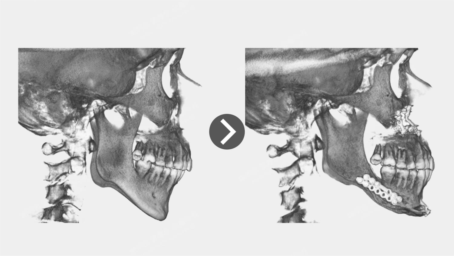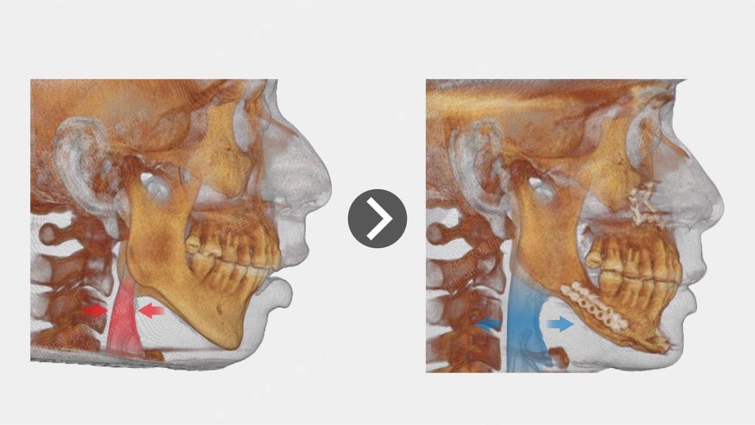




| 術前症狀 | 手術計畫 |
| 夜間嚴重打鼾與日間嗜睡,中度睡眠呼吸中止症(AHI 17.3) | 進行逆時針旋轉型雙顎前移手術,前移上顎、下顎與舌骨以拓寬氣道 |
診療計畫及術後改變
這位患者主訴為夜間嚴重打鼾及日間嗜睡,影響生活品質。多項睡眠檢查結果顯示中度阻塞性睡眠呼吸中止症(AHI = 17.3),睡眠期間最低血氧濃度降至 83%,顯示顯著的通氣障礙。
This patient presented with complaints of severe snoring during sleep and excessive daytime sleepiness, significantly affecting quality of life. Polysomnography revealed moderate obstructive sleep apnea (OSA) with an apnea–hypopnea index (AHI) of 17.3, and a minimum oxygen saturation of 83% during sleep, indicating substantial ventilatory compromise.
電腦斷層影像顯示,患者後方呼吸道空間明顯狹窄,主因為上下顎骨發育不足,未能適當向前成長。上顎骨後縮合併氣道塌陷,使鬆弛的懸壅垂在狹窄空間中震動,產生明顯鼾聲;下顎骨後縮則導致舌頭後傾、舌肌張力不足、舌骨位置偏低,成為阻塞性呼吸中止的主要結構性因素。
Computed tomography (CT) imaging demonstrated a markedly narrowed posterior airway space, primarily due to underdeveloped maxillary and mandibular growth. The retruded maxilla, combined with airway collapse, allowed the relaxed uvula to vibrate within the constricted space, producing loud snoring. The retrognathic mandible further contributed to posterior tongue displacement, reduced tongue muscle tone, and a low-positioned hyoid bone—key anatomical factors in the pathogenesis of OSA.
針對上述解剖與功能問題,我們為患者規劃逆時針旋轉型雙顎前移手術(Counter-Clockwise Rotation Maxillomandibular Advancement, CCWR-MMA)。
整個手術設計的中心思想有三個:
1. 上顎骨前移有效拉緊軟顎肌群,使懸壅垂向前牽引,遠離後咽壁,減少鼾聲產生
2. 下顎骨前移則牽引舌根部位,使舌頭向前移動,減少舌根後墜
3. 與此同時,舌骨亦被牽引至前上方位置,遠離氣道後壁,顯著擴大咽腔空間,改善通氣效率。
To address these structural and functional issues, we planned a counter-clockwise rotation maxillomandibular advancement (CCWR-MMA) procedure. This surgical approach included:
- Maxillary advancement, which tightened the soft palate musculature and repositioned the uvula anteriorly, away from the posterior pharyngeal wall, thereby reducing snoring;
- Mandibular advancement, which, through its anatomical connection to the tongue base, repositioned the tongue anteriorly, minimizing posterior collapse;
- Additionally, the hyoid bone was drawn forward and upward, distancing it from the posterior airway wall and significantly expanding the pharyngeal space to improve airflow.
術前與術後的電腦斷層影像清楚顯示,骨骼位置調整後,軟組織張力提升,氣道空間隨之拓寬,結構性改善明確。
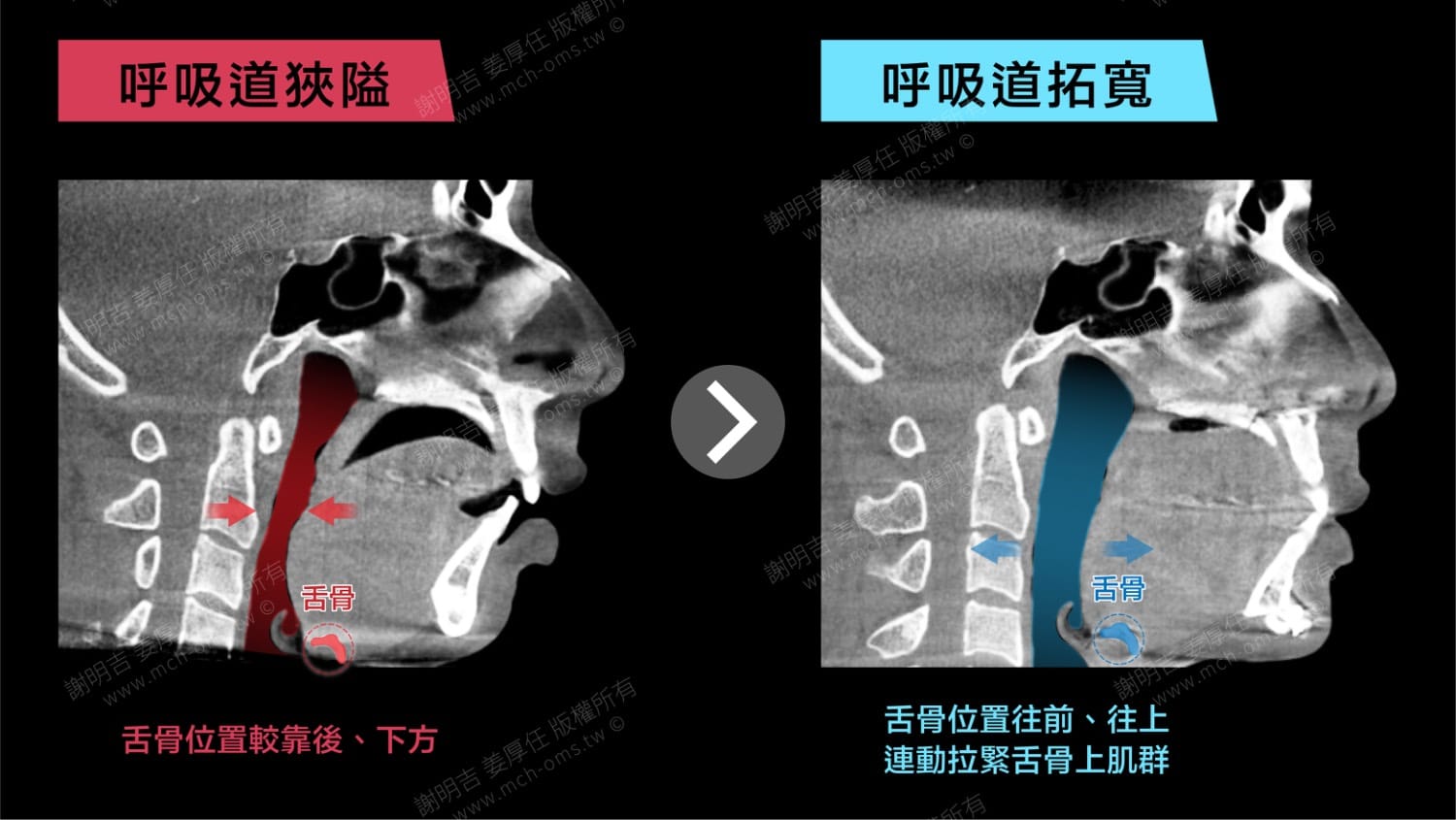
術後睡眠報告顯示 AHI 降至 6.3,最低血氧濃度提升至 89%。患者術後自述睡眠品質顯著改善,日間精神狀態明朗,整體生活品質亦獲得提升。
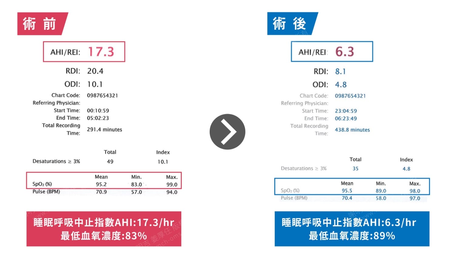
Pre- and postoperative CT scans clearly demonstrated how skeletal repositioning led to soft tissue tensioning and airway enlargement. Postoperatively, the patient’s AHI decreased from 17.3 to 6.3, and minimum oxygen saturation improved from 83% to 89%. Clinically, the patient reported markedly improved sleep quality and daytime alertness, with a refreshed and energized demeanor.
外觀改變


手術前後斷層掃描對比